“
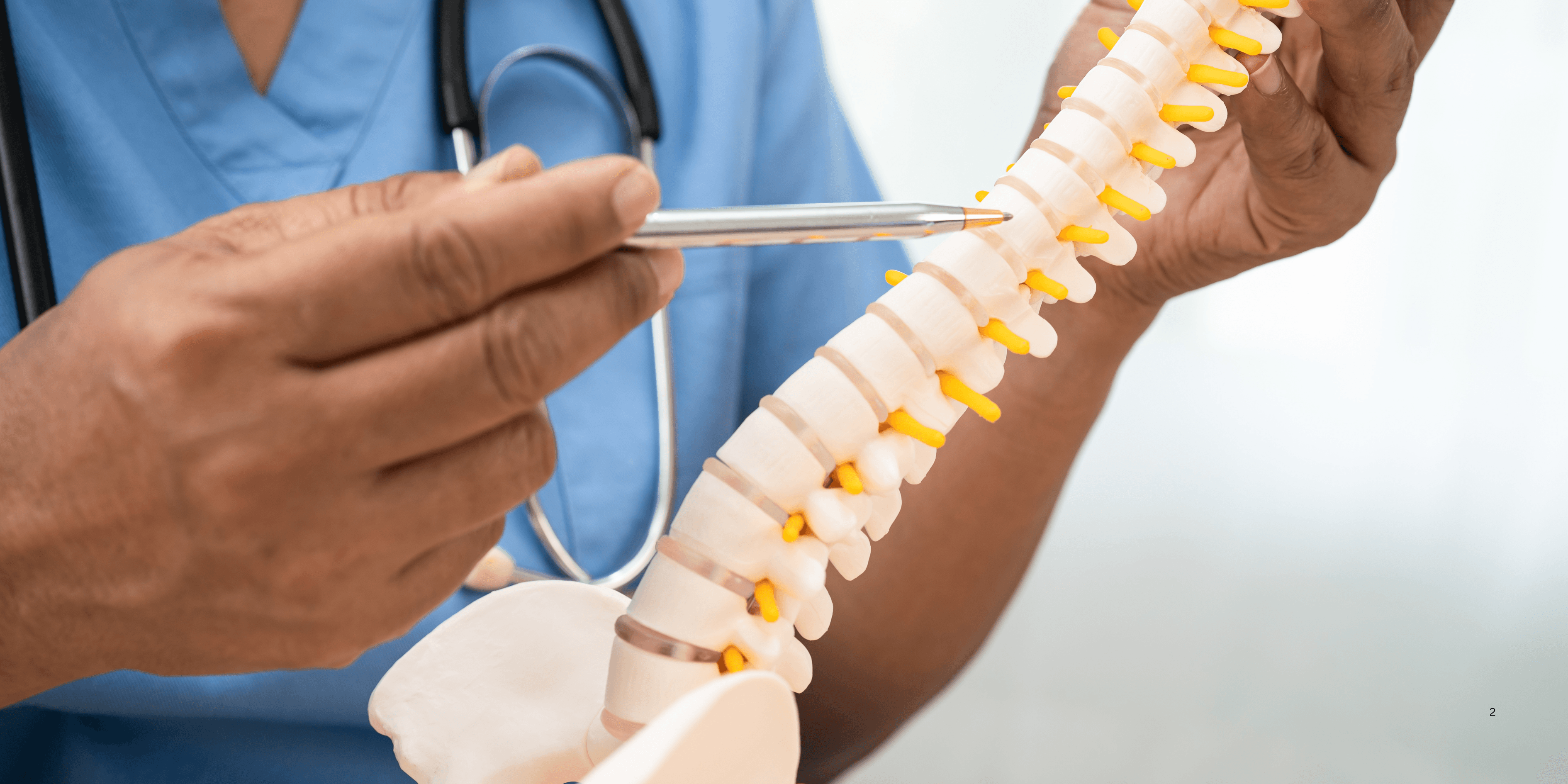
The structure of the spinal cord plays a vital role in connecting the brain with the rest of the body, acting as the core highway for nerve signals. Protected by the vertebral column, the spinal cord is organized into segments, nerves, and supporting tissues that coordinate sensation and movement. This remarkable structure contains both gray and white matter, shaped like a butterfly in cross-section, and is crucial in everything from reflexes to complex motor control. 1
1
”
Greek physician Galen, one of history’s early anatomists, identified the spinal cord as a continuation of the brain, describing its tubular structure and noting its crucial role in movement and sensation.1
The spinal cord extends from the brainstem to the lower back, specifically ending near the first or second lumbar vertebra, forming a tapered point known as the conus medullaris. 2
It is encased within the spinal canal and shielded by vertebrae, cerebrospinal fluid, and meninges, which serve as physical and chemical protection against impact and infection. 3
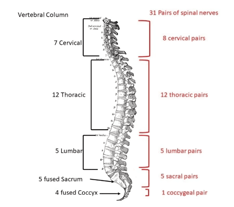
The spinal cord consists of 31 segments, each giving rise to a pair of spinal nerves—these nerves emerge through spaces between the vertebrae and connect to different body regions.
Each segment is named based on the vertebrae it aligns with—cervical, thoracic, lumbar, sacral, and coccygeal—and controls specific motor and sensory functions. 4
Inside, the spinal cord is divided into gray matter, shaped like a butterfly, and white matter, which surrounds it; the gray matter processes information, while the white matter carries messages. 5
The gray matter houses motor neurons, interneurons, and sensory neurons, enabling reflexes and basic motor responses independent of the brain. 6
White matter contains myelinated axons arranged in ascending and descending tracts, which conduct sensory data to the brain and motor commands from it. 7
The central canal, a tiny fluid-filled space running through the spinal cord’s core, carries cerebrospinal fluid and helps maintain internal chemical balance and cushioning. 8
The dorsal roots of spinal nerves carry sensory information into the spinal cord, while the ventral roots transmit motor commands from the spinal cord to muscles. 9
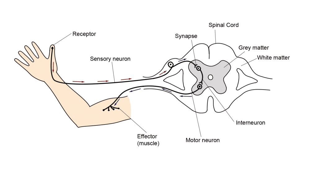
Reflex arcs—like pulling your hand away from a hot object—are processed directly in the spinal cord without needing input from the brain, allowing fast reactions.
The lumbar enlargement, located between T9 and T12, gives rise to nerves that serve the pelvis and legs, supporting standing, walking, and leg coordination. 10
The cauda equina, a bundle of spinal nerves and nerve roots resembling a horse’s tail, extends beyond the conus medullaris and controls lower body functions. 11
The spinal cord grows more slowly than the vertebral column, so in adults, the spinal cord ends higher than in newborns, where it typically reaches into the lower lumbar area. 12
Blood supply to the spinal cord comes from the anterior spinal artery and paired posterior spinal arteries, which branch from the vertebral arteries and radicular arteries. 13
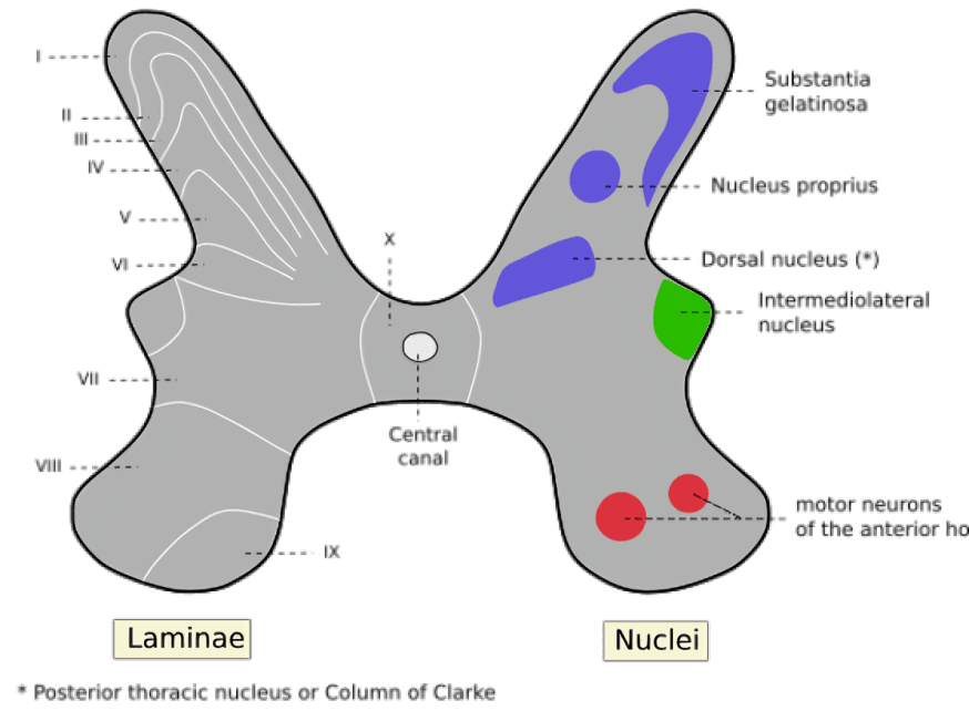
Neurons in the spinal cord are organized into laminae—ten distinct layers that perform specific processing roles related to pain, touch, proprioception, and motor output.
The spinal cord’s segmental organization ensures that injury at any point leads to specific motor and sensory deficits, aiding doctors in diagnosing neurological damage. 14
Ependymal cells lining the central canal play a role in producing cerebrospinal fluid, while astrocytes and microglia provide structural and immune support within the spinal tissue. 15
In cross-section, the butterfly-shaped gray matter is divided into dorsal horns (sensory), ventral horns (motor), and lateral horns (autonomic), each managing different signal types. 16
The concept of neural pathways in the spinal cord has inspired treatments like spinal cord stimulation, which uses electrical impulses to relieve pain or restore lost motor control.17