“
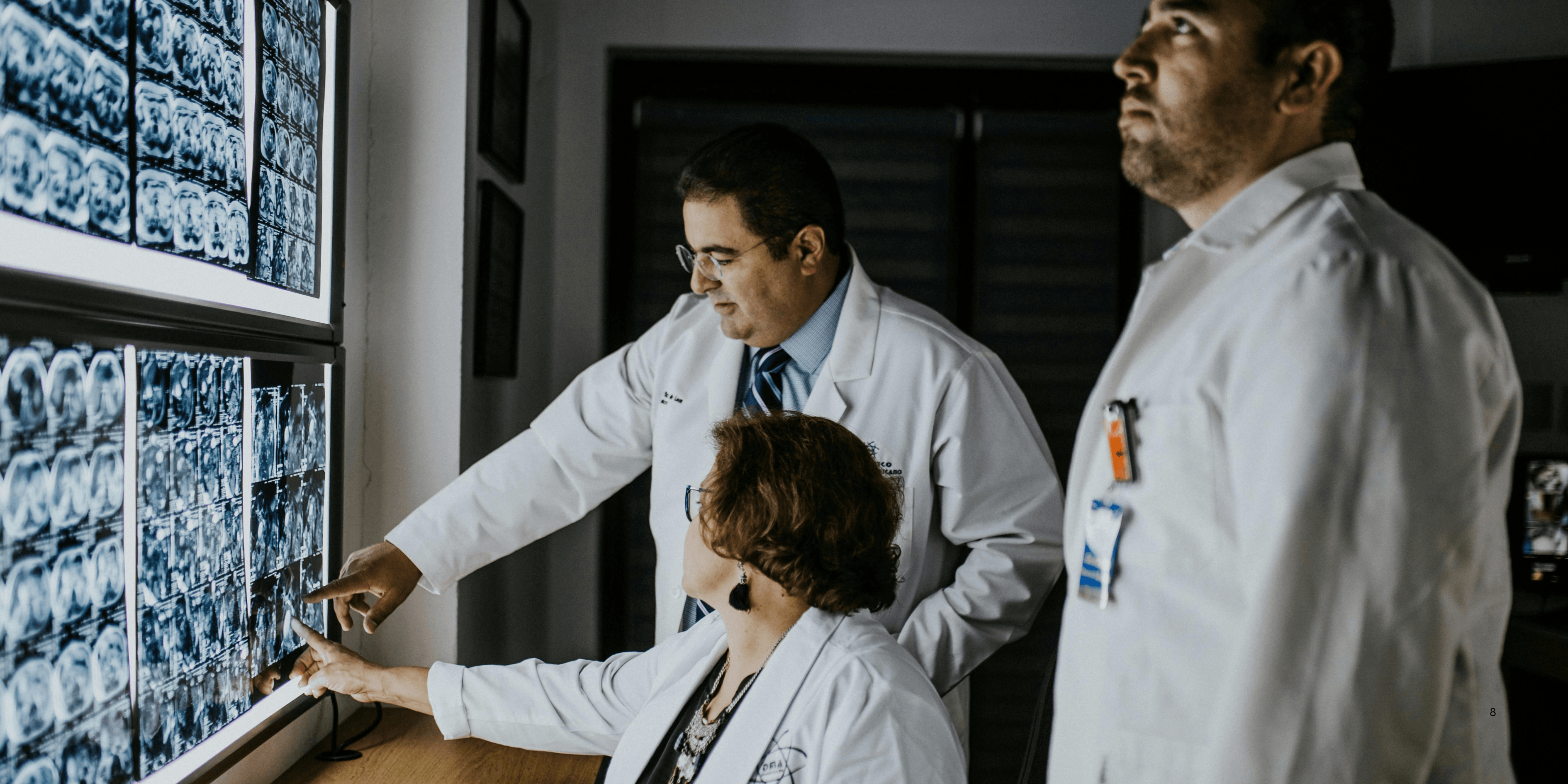
When diagnosing brain conditions, two major imaging tools dominate medical practice: Brain Scans: MRI vs. CT. Each scan serves a vital purpose in revealing different aspects of brain structure, injury, and disease. MRIs use powerful magnets and radio waves, while CT scans utilize X-ray technology.1
1
”
CT scans use ionizing radiation to generate detailed images of the brain in minutes, making them ideal for identifying skull fractures, strokes, or bleeding in emergency situations quickly. 1
Unlike CT scans, MRIs don’t expose patients to radiation, which is especially beneficial for repeated scanning or for vulnerable groups such as children, pregnant women, or patients with radiation sensitivities. 2
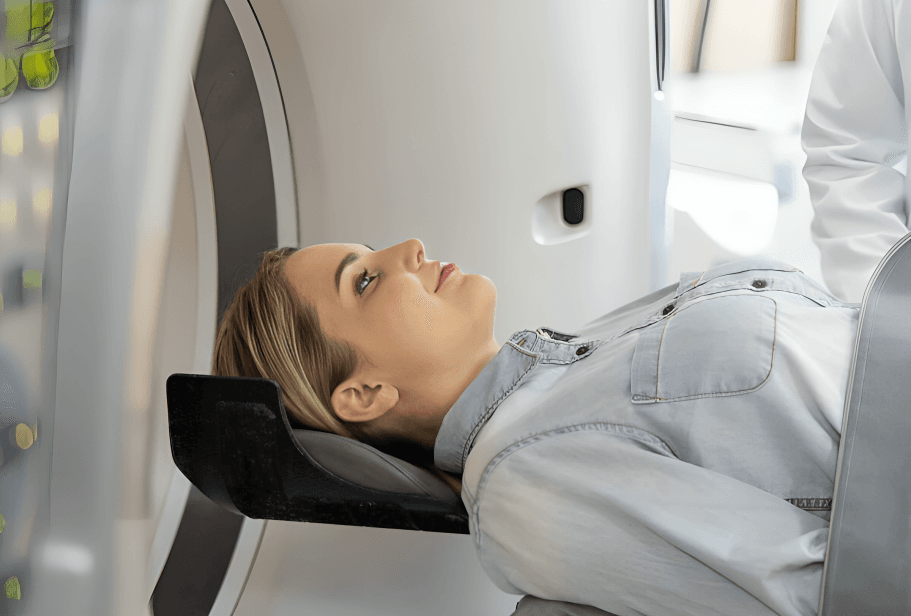
An MRI of the brain takes longer than a CT—typically 30 to 60 minutes compared to a CT’s 5 to 10—making CT more practical for emergencies where time is a critical factor in diagnosis.
Patients with metal implants, such as pacemakers or cochlear devices, may be advised against MRI scans, as the strong magnetic field can interfere with or move these metallic objects dangerously. 3
CT scans excel in identifying acute head trauma and bone fractures, while MRIs are superior for examining soft tissue, white matter changes, and conditions like multiple sclerosis or brain infections. 4
Some CT scans use a contrast dye (iodine-based), while MRIs use gadolinium. Both contrast agents help highlight abnormalities, but gadolinium is generally considered safer with fewer allergic reactions. 5
A brain CT is often the first step in stroke evaluation to rule out bleeding, followed by MRI to detect ischemic (non-bleeding) strokes that may not initially show up in CT images. 6
MRIs can detect demyelinating diseases like multiple sclerosis more clearly than CTs due to their superior sensitivity to changes in white matter and subtle nerve tissue abnormalities. 7
Claustrophobia is more common during MRI scans because the patient must lie still inside a narrow tube for extended periods, whereas CT scans are faster and less enclosed in design. 8

CT scans are widely available in smaller hospitals and clinics, while MRI machines are less common due to their higher cost, space requirements, and technical complexity in operation.
In neurology, MRIs are preferred for diagnosing brain tumors, as their high contrast resolution reveals tumor boundaries, surrounding edema, and pressure effects on brain structures more precisely. 9
A single CT scan exposes a person to a level of radiation equivalent to several hundred chest X-rays, raising concerns about cumulative radiation risks with repeated use over time. 10
For detecting hemorrhages, skull fractures, or swelling after a traumatic brain injury, CT is typically used first due to its speed, accessibility, and ability to capture bone and blood density differences. 11
Functional MRI (fMRI), a specialized type of brain MRI, helps researchers and doctors study brain activity by measuring changes in blood flow during tasks, such as speaking, thinking, or moving. 12
Pediatric brain scanning favors MRI over CT to avoid radiation exposure in growing brains, though sedation may be needed for younger children due to the longer duration and noise levels of MRI machines. 13
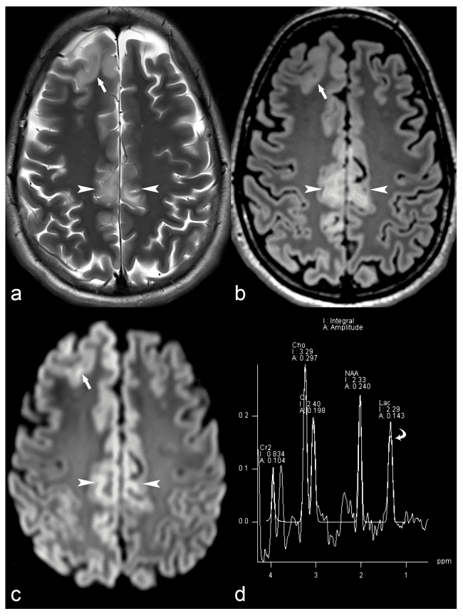
CT scans may miss smaller lesions, making MRI the better choice for conditions like epilepsy, neurodegenerative disorders, and brain infections requiring high-detail imaging of internal structures.
For surgical planning, especially in brain tumor removal or epilepsy treatment, MRI provides 3D anatomical maps that guide neurosurgeons in avoiding critical areas and preserving brain function. 14
MRI provides multi-plane views (sagittal, coronal, and axial) without repositioning the patient, allowing doctors to view the brain from different angles with clarity unmatched by standard CT technology. 15
MRI scans often reveal more incidental findings—unexpected abnormalities not related to symptoms—which may lead to additional tests, while CT scans usually highlight only major visible issues. 16
Philosopher and mathematician René Descartes once described the brain as the "seat of the soul"; modern doctors now rely on brain scans (MRI vs. CT) to explore that seat with science instead of speculation. 17